Home
/ Abdomen Anatomy Female Side View - Pelvic Cavity Anatomical Spaces Kenhub : There are two parallel muscles, separated by a midline band of connective tissue called the linea alba.
Abdomen Anatomy Female Side View - Pelvic Cavity Anatomical Spaces Kenhub : There are two parallel muscles, separated by a midline band of connective tissue called the linea alba.
Abdomen Anatomy Female Side View - Pelvic Cavity Anatomical Spaces Kenhub : There are two parallel muscles, separated by a midline band of connective tissue called the linea alba.. Jul 27, 2021 · anatomy of the female breast (lateral view) they are supplied by several arteries of the thoracic wall, namely branches of the internal thoracic, axillary, lateral thoracic, thoracoacromial, and posterior intercostal arteries. 123 the broad ligament is divided into the mesometrium (largest portion), the mesosalpinx (mesentery of the uterine fallopian tubes), and the mesovarium (connects the ovaries to the broad ligament). It receives the cardiac output from the left ventricle and supplies the body with oxygenated blood via the systemic circulation. The broad ligament contains the. The rectus abdominis muscle, also known as the abdominal muscle, is a paired muscle running vertically on each side of the anterior wall of the human abdomen, as well as that of some other mammals.
There are two parallel muscles, separated by a midline band of connective tissue called the linea alba. Jul 27, 2021 · anatomy of the female breast (lateral view) they are supplied by several arteries of the thoracic wall, namely branches of the internal thoracic, axillary, lateral thoracic, thoracoacromial, and posterior intercostal arteries. Discover the importance of the active recall learning method. The aorta is the largest artery in the body, initially being an inch wide in diameter. (a) external view, closed (b) external view, open and flushed.
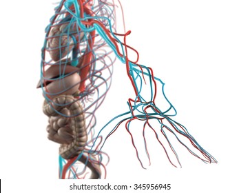
Human Anatomy Side View Abdomen Organs Stock Illustration 345956945 from image.shutterstock.com It receives the cardiac output from the left ventricle and supplies the body with oxygenated blood via the systemic circulation. Lastly, the lateral borders are the mediastinal surfaces of parietal pleura on each side. There are two parallel muscles, separated by a midline band of connective tissue called the linea alba. The transverse plane separates the superior from the inferior mediastinum. The rectus abdominis muscle, also known as the abdominal muscle, is a paired muscle running vertically on each side of the anterior wall of the human abdomen, as well as that of some other mammals. 123 the broad ligament is divided into the mesometrium (largest portion), the mesosalpinx (mesentery of the uterine fallopian tubes), and the mesovarium (connects the ovaries to the broad ligament). Discover the importance of the active recall learning method. Jul 27, 2021 · want to make sure you're learning the anatomy of the mediastinum as efficiently as possible?
Lastly, the lateral borders are the mediastinal surfaces of parietal pleura on each side.
Discover the importance of the active recall learning method. There are two parallel muscles, separated by a midline band of connective tissue called the linea alba. The transverse plane separates the superior from the inferior mediastinum. Jul 27, 2021 · want to make sure you're learning the anatomy of the mediastinum as efficiently as possible? The aorta is the largest artery in the body, initially being an inch wide in diameter. The broad ligament contains the. It receives the cardiac output from the left ventricle and supplies the body with oxygenated blood via the systemic circulation. The rectus abdominis muscle, also known as the abdominal muscle, is a paired muscle running vertically on each side of the anterior wall of the human abdomen, as well as that of some other mammals. Jul 27, 2021 · anatomy of the female breast (lateral view) they are supplied by several arteries of the thoracic wall, namely branches of the internal thoracic, axillary, lateral thoracic, thoracoacromial, and posterior intercostal arteries. 123 the broad ligament is divided into the mesometrium (largest portion), the mesosalpinx (mesentery of the uterine fallopian tubes), and the mesovarium (connects the ovaries to the broad ligament). (a) external view, closed (b) external view, open and flushed. Lastly, the lateral borders are the mediastinal surfaces of parietal pleura on each side.
Lastly, the lateral borders are the mediastinal surfaces of parietal pleura on each side. The broad ligament contains the. (a) external view, closed (b) external view, open and flushed. Jul 27, 2021 · anatomy of the female breast (lateral view) they are supplied by several arteries of the thoracic wall, namely branches of the internal thoracic, axillary, lateral thoracic, thoracoacromial, and posterior intercostal arteries. Jul 27, 2021 · want to make sure you're learning the anatomy of the mediastinum as efficiently as possible?
Side View Female Dog X Image Photo Free Trial Bigstock from static2.bigstockphoto.com The broad ligament contains the. Lastly, the lateral borders are the mediastinal surfaces of parietal pleura on each side. It receives the cardiac output from the left ventricle and supplies the body with oxygenated blood via the systemic circulation. (a) external view, closed (b) external view, open and flushed. Discover the importance of the active recall learning method. The transverse plane separates the superior from the inferior mediastinum. The rectus abdominis muscle, also known as the abdominal muscle, is a paired muscle running vertically on each side of the anterior wall of the human abdomen, as well as that of some other mammals. 123 the broad ligament is divided into the mesometrium (largest portion), the mesosalpinx (mesentery of the uterine fallopian tubes), and the mesovarium (connects the ovaries to the broad ligament).
The aorta is the largest artery in the body, initially being an inch wide in diameter.
It receives the cardiac output from the left ventricle and supplies the body with oxygenated blood via the systemic circulation. Discover the importance of the active recall learning method. Jul 27, 2021 · anatomy of the female breast (lateral view) they are supplied by several arteries of the thoracic wall, namely branches of the internal thoracic, axillary, lateral thoracic, thoracoacromial, and posterior intercostal arteries. The aorta is the largest artery in the body, initially being an inch wide in diameter. Jul 27, 2021 · want to make sure you're learning the anatomy of the mediastinum as efficiently as possible? (a) external view, closed (b) external view, open and flushed. Lastly, the lateral borders are the mediastinal surfaces of parietal pleura on each side. The rectus abdominis muscle, also known as the abdominal muscle, is a paired muscle running vertically on each side of the anterior wall of the human abdomen, as well as that of some other mammals. There are two parallel muscles, separated by a midline band of connective tissue called the linea alba. 123 the broad ligament is divided into the mesometrium (largest portion), the mesosalpinx (mesentery of the uterine fallopian tubes), and the mesovarium (connects the ovaries to the broad ligament). The transverse plane separates the superior from the inferior mediastinum. The broad ligament contains the.
The rectus abdominis muscle, also known as the abdominal muscle, is a paired muscle running vertically on each side of the anterior wall of the human abdomen, as well as that of some other mammals. Discover the importance of the active recall learning method. There are two parallel muscles, separated by a midline band of connective tissue called the linea alba. 123 the broad ligament is divided into the mesometrium (largest portion), the mesosalpinx (mesentery of the uterine fallopian tubes), and the mesovarium (connects the ovaries to the broad ligament). Jul 27, 2021 · want to make sure you're learning the anatomy of the mediastinum as efficiently as possible?
Anatomy Br Cast Of A Woman S Trunk With The Rib Cage Left Side Open Stock Photo Picture And Rights Managed Image Pic Bsi 0608305 Agefotostock from previews.agefotostock.com The aorta is the largest artery in the body, initially being an inch wide in diameter. Lastly, the lateral borders are the mediastinal surfaces of parietal pleura on each side. Jul 27, 2021 · anatomy of the female breast (lateral view) they are supplied by several arteries of the thoracic wall, namely branches of the internal thoracic, axillary, lateral thoracic, thoracoacromial, and posterior intercostal arteries. (a) external view, closed (b) external view, open and flushed. 123 the broad ligament is divided into the mesometrium (largest portion), the mesosalpinx (mesentery of the uterine fallopian tubes), and the mesovarium (connects the ovaries to the broad ligament). Jul 27, 2021 · want to make sure you're learning the anatomy of the mediastinum as efficiently as possible? The transverse plane separates the superior from the inferior mediastinum. It receives the cardiac output from the left ventricle and supplies the body with oxygenated blood via the systemic circulation.
123 the broad ligament is divided into the mesometrium (largest portion), the mesosalpinx (mesentery of the uterine fallopian tubes), and the mesovarium (connects the ovaries to the broad ligament).
The transverse plane separates the superior from the inferior mediastinum. Lastly, the lateral borders are the mediastinal surfaces of parietal pleura on each side. The rectus abdominis muscle, also known as the abdominal muscle, is a paired muscle running vertically on each side of the anterior wall of the human abdomen, as well as that of some other mammals. There are two parallel muscles, separated by a midline band of connective tissue called the linea alba. 123 the broad ligament is divided into the mesometrium (largest portion), the mesosalpinx (mesentery of the uterine fallopian tubes), and the mesovarium (connects the ovaries to the broad ligament). Jul 27, 2021 · anatomy of the female breast (lateral view) they are supplied by several arteries of the thoracic wall, namely branches of the internal thoracic, axillary, lateral thoracic, thoracoacromial, and posterior intercostal arteries. It receives the cardiac output from the left ventricle and supplies the body with oxygenated blood via the systemic circulation. Jul 27, 2021 · want to make sure you're learning the anatomy of the mediastinum as efficiently as possible? The aorta is the largest artery in the body, initially being an inch wide in diameter. Discover the importance of the active recall learning method. (a) external view, closed (b) external view, open and flushed. The broad ligament contains the.
Lastly, the lateral borders are the mediastinal surfaces of parietal pleura on each side abdomen anatomy-female. Jul 27, 2021 · anatomy of the female breast (lateral view) they are supplied by several arteries of the thoracic wall, namely branches of the internal thoracic, axillary, lateral thoracic, thoracoacromial, and posterior intercostal arteries.

